Light microscopy(LM)
1.觀看順序#
- LPF(看cortex, medulla比例, 是否有其他tissue, IFTA, interstitial inflammation/infiltrations)
- HPF
- Glomerulus
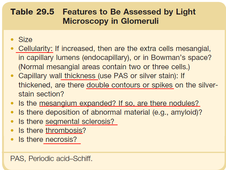
Table 29.5 Features to Be Assessed by Light Microscopy in Glomeruli#
- Size
- Cellularity: If increased, then are the extra cells mesangial, in capillary lumens (endocapillary), or in Bowman's space? (Normal mesangial areas contain two or three cells.)
- Capillary wall thickness (use PAS or silver stain): If thickened, are there double contours or spikes on the silver-stain section?
- Is the mesangium expanded? If so, are there nodules?
- Is there deposition of abnormal material (e.g., amyloid)?
- Is there segmental sclerosis?
- Is there thrombosis?
- Is there necrosis?
PAS, Periodic acid–Schiff.
- Thrombus, cells*4(WBC, mesangial cells, podocytes, crescent), thickness/double contour/spikes, matrix(nodules?), sclerosis? necrosis?
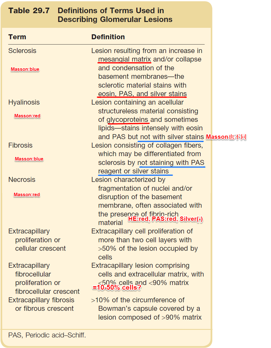
Table 29.7 Definitions of Terms Used in Describing Glomerular Lesions#
| Term | Definition |
|---|---|
| Sclerosis | Lesion resulting from an increase in mesangial matrix and/or collapse and condensation of the basement membranes—the sclerotic material stains with eosin, PAS, and silver stains. Masson:blue |
| Hyalinosis | Lesion containing an acellular structureless material consisting of glycoproteins and sometimes lipids—stains intensely with eosin and PAS but not with silver stains. Masson:red (Masson也不同) |
| Fibrosis | Lesion consisting of collagen fibers, which may be differentiated from sclerosis by not staining with PAS reagent or silver stains. Masson:blue |
| Necrosis | Lesion characterized by fragmentation of nuclei and/or disruption of the basement membrane, often associated with the presence of fibrin-rich material. HE:red, PAS:red, Silver(-) |
| Extracapillary proliferation or cellular crescent | Extracapillary cell proliferation of more than two cell layers with >50% of the lesion occupied by cells |
| Fibrocellular proliferation or fibrocellular crescent | cells and extracellular matrix, with 10-50% cells 且 >50%~<90% matrix(對比: cellular >50% cells; fibrous <10% cells 且 >90% matrix) |
| Extracapillary fibrosis or fibrous crescent | >10% of the circumference of Bowman's capsule covered by a lesion composed of >90% matrix |
PAS, Periodic acid–Schiff.
- Tubules
- Percentage atrophy
- Signs of acute damage (e.g., Lumen dilatation, epithelial flattening, granular casts, mitoses)
- Tubulitis
- Casts: Granular casts suggest acute tubular injury; eosinophilic fractured casts suggest myeloma (Myeloma cast比較硬); neutrophil casts suggest acute pyelonephritis
- Crystals (e.g., oxalate)
- Viral inclusions (e.g., BK virus)
- Tubules, vacuolization, inclusions, WBCs, deposits/crystals/casts
- Interstitium
- BV
- Thrombosis? embolism? intimal thickening? hyalinosis? reduplication of internal elastic lamina? medial hyperplasia? necrosis? inflammation?…
2.Normal glomerulus#
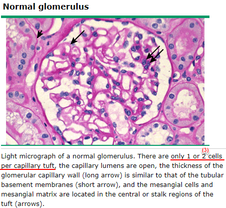
Light micrograph of a normal glomerulus. There are only 1 or 2 cells per capillary tuft, the capillary lumens are open, the thickness of the glomerular capillary wall (long arrow) is similar to that of the tubular basement membranes (short arrow), and the mesangial cells and mesangial matrix are located in the central or stalk regions of the tuft (arrows).
- Endothelium和mesangial cells會靠近集中
- Stains
- 1.PAS stain(=Periodic Acid-Schiff stain), 用過碘酸把醣類相鄰兩個碳上的OH基氧化成醛基, 再用Schiff試劑和醛基反應使呈現紫紅色
- 會將Bowman's capsule, mesangial matrix, basement membrane(BM)染成紫紅色所以可清楚看出tubular atrophy and crescent
- 也會染sclerosis, hyaline and T-H proteins
- 2.Grocott's methenamine silver stain(=Chromic Acid Silver Methenamine(CSM)): 染DNA and proteins(collagens, reticulins..., 所以可染Bowman's capsule, mesangial matrix, basement membrane(BM)...)
- 常用silver stain看有否double contour of GBM(明顯的其他stain也看得到) and spikes(主要是因為silver染的東西比較少但其中包含GBM, 所以特別好看GBM)
- Double contour of GBM有很多種成因
- MGN:GBM去包住subepithelial depositions, 形成兩層的樣貌(中間是deposition, 所以兩層都是GBM), 有spikes
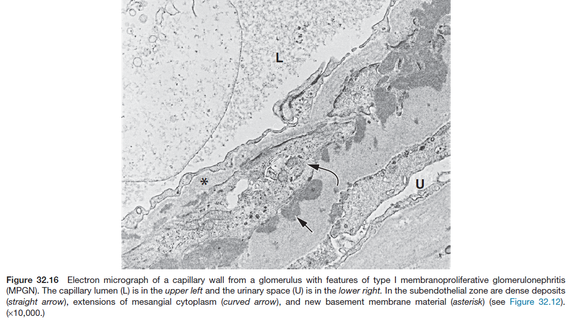
- MPGN:Mesangial cells去包住endothelium(為了隔開subendothelial depositions; 叫mesangial interposition), 形成兩層的樣貌(內層是新形成的GBM(介於endothelium和mesangial interposition間), 中間是mesangial包過來的部分, 外層是subendothelial depositions+原GBM), 沒有spikes
- Type II or (type III)(type III應該大多還是MPGN pattern吧?)不一定有MPGN pattern
- 因為DDD(=type II)是intramembranous dense depositions直接將GBM分成兩層, 所以不一定看得到
- Chronic transplant glomerulopathy(=chronic ABMR), LN type III/IV(本來就是MPGN的D/D), cryoglobulinemia(本來就是MPGN pattern), IgAN/HSP(比較嚴重可以MPGN pattern), PIGN/IRGN(本來就是MPGN pattern), MIDD(大多是nodular pattern, 但是可以MPGN pattern)/fibrillary GN(15%是MPGN pattern)/immunotactoid GN(大多為MPGN pattern)/collagenofibrotic glomerulopathy也可以有double contour(MPGN pattern)
- Chronic transplant glomerulopathy, LN III/IV, 感染類, 某些nodular pattern類可double contour
- Type II or (type III)(type III應該大多還是MPGN pattern吧?)不一定有MPGN pattern
- Chronic TMA:一樣是mesangial interposition(同MPGN), 中間沒有deposits也沒有spikes
- 本來chronic TMA就會MPGN pattern...; 所以要看EM, immunofluorescence才能和MPGN分
- 1.PAS stain(=Periodic Acid-Schiff stain), 用過碘酸把醣類相鄰兩個碳上的OH基氧化成醛基, 再用Schiff試劑和醛基反應使呈現紫紅色
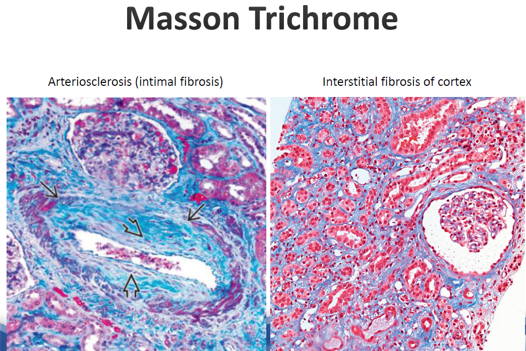
- Masson trichrome stain
- 一樣染Bowman's capsule, mesangial matrix, basement membrane(BM)..., 主要用來看interstitial fibrosis
- 一般mesangial matrix/sclerosis是藍色的, 但immunocomplex, fibrin(necrosis)是紅色的, 所以可以協助看immunocomplex depositions...
- 一樣染Bowman's capsule, mesangial matrix, basement membrane(BM)..., 主要用來看interstitial fibrosis
3.Glomeruli(須至少看>20 glomerulus才比較能exclude mild, focal glomerular disease; 至少也要>10 glomerulus)(林維洲醫師希望medulla佔切片的1/3左右, 主要是FSGS多在medulla area...)#
- 8種主要glomerular patterns
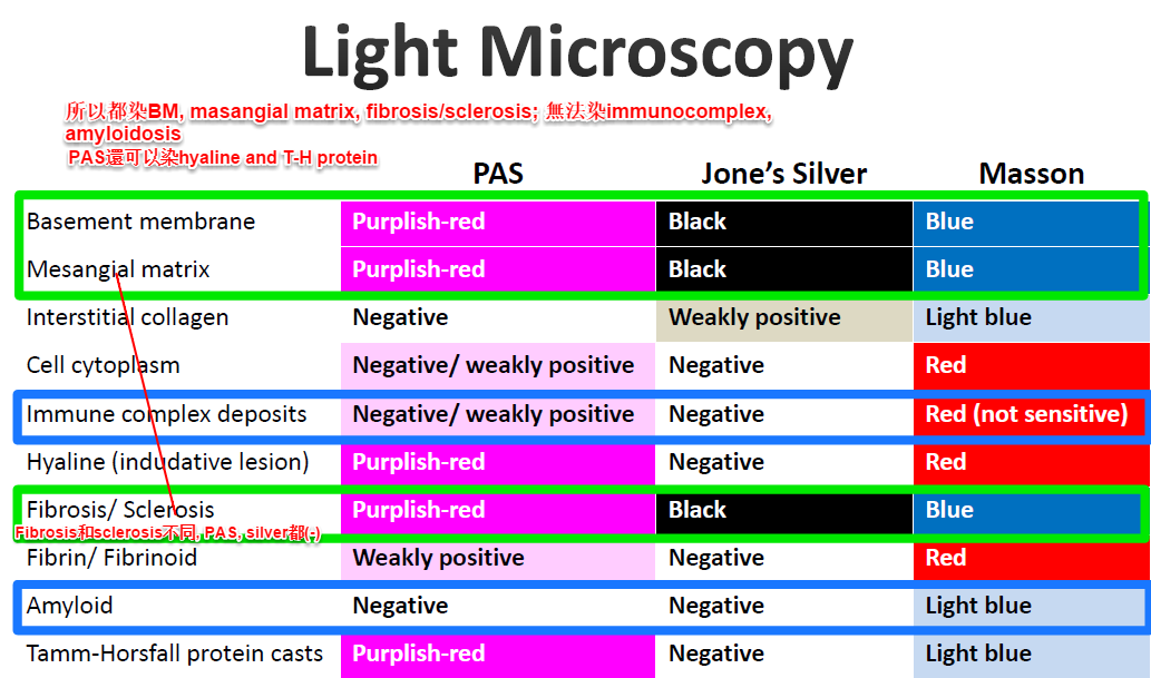
Table: Light Microscopy Staining Summary#
| PAS | Jone's Silver | Masson | |
|---|---|---|---|
| Basement membrane | Purple-red | Black | Blue |
| Mesangial matrix | Purple-red | Black | Blue |
| Cell cytoplasm | Negative | Weakly positive | Red |
| Immune complex deposits | Purple | Negative | Red (not sensitive) |
| Hyaline (indicative lesion) | Purple-red | Negative | Red |
| Fibrosis/ Sclerosis | Purple-red | Black | Blue |
| Fibrinoid (necrosis) | Weakly positive | Negative | Red |
| Amyloid | Negative | Negative | Light blue |
| Tamm-Horsfall protein casts | Purple-red | Negative | Light blue |
- BM, mesangial matrix, sclerosis在PAS, silver, Masson trichrome都(+)
- PAS也染hyaline和T-H protein(所以cast(+))
- Silver其他東西都不太染
- Masson trichrome stain的cell cytoplasm, hyaline, fibrinoid(necrosis), immunocomplex是紅色的(如MIDD, 但amyloidosis是gray-purple)
- 其他也可染C4d(ABMR), IgG subclass(MIDD...), simian virus 40(SV40) large T antigen(BK, JC virus), EBV/CMV Ag, congo red(amyloidosis; 需要厚切(10um厚)), serum amyloid A(AA), type IV collagen的α3, α5 chains(TBMD, Alport's syndrome), type III collagen(collagenofibrotic glomerulopathy), fibronectin(fibronectin glomerulopathy), von Kossa stain(染Ca), Perls Prussian blue stain(染iron), PLA2R(MGN), THSD7A(MGN);新抗原 EXT1/EXT2, NELL1, SEMA3B, PCDH7, HTRA1, NCAM1(各有不同臨床病理)〔PMID 36016931〕, myoglobin, lymphocyte surface Ag(lymphoma...)
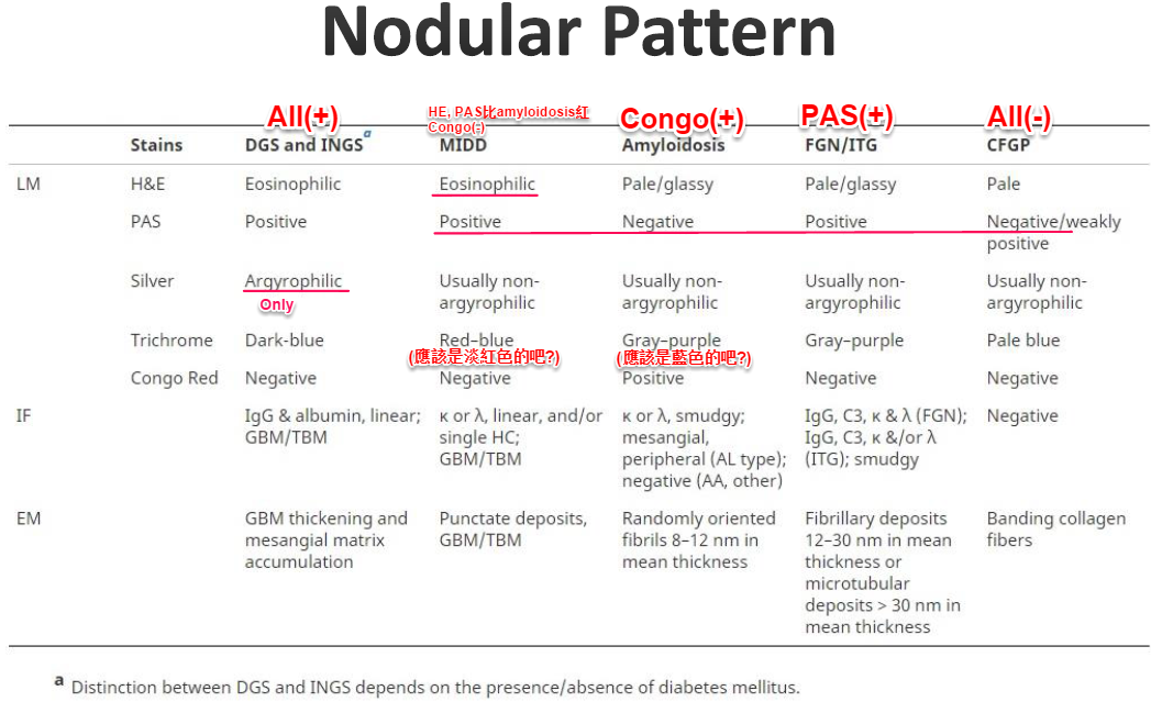
Nodular Pattern - Differential Diagnosis Table#
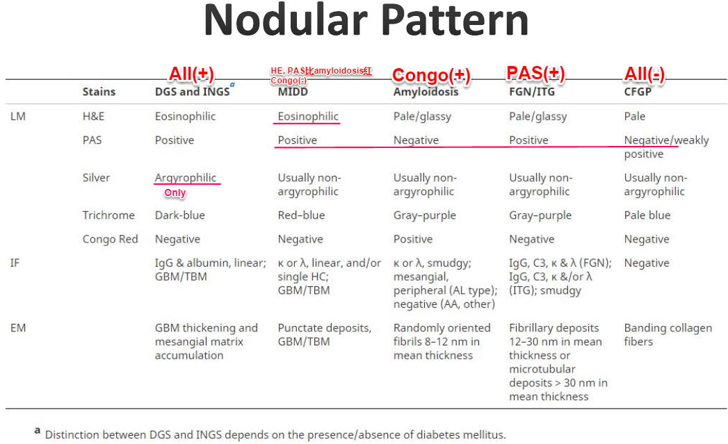
【結節性病變秒殺關鍵】(Brenner 11e Ch 29): 五大結節性腎絲球病變中,唯有糖尿病腎硬化症 (DN / K-W nodules) 在 Jones' Silver 染色呈現 Argyrophilic (黑色網狀陽性),其餘 Amyloidosis、MIDD、Fibrillary GN、Collagenofibrotic GP 之結節在銀染均呈陰性 (Usually non-argyrophilic)!
| 染色特徵 | DGS and INGS(H&E+、PAS+、Silver+) | MIDD(H&E+、PAS+、Congo−) | Amyloidosis(Congo+) | FGN/ITG(PAS+、Congo−) | CFGP(All−) |
|---|---|---|---|---|---|
| LM H&E | Eosinophilic | Eosinophilic | Pale/glassy | Pale/glassy | Pale |
| LM PAS | Positive | Positive | Negative | Positive | Negative/weakly positive |
| LM Silver | Argyrophilic Only | Usually non-argyrophilic | Usually non-argyrophilic | Usually non-argyrophilic | Usually non-argyrophilic |
| LM Trichrome | Dark-blue | Red-blue(MIDD 為 fuchsinophilic 淡紅/紫紅) | Gray-purple(amyloid 為灰紫/淡藍) | Gray-purple | Pale blue |
| LM Congo Red | Negative | Negative | Positive | Negative | Negative |
| IF | IgG & albumin, linear; GBM/TBM | κ or λ, linear, and/or single HC; GBM/TBM | κ or λ, smudgy; mesangial, peripheral (AL type); negative (AA, other) | IgG, C3, κ & λ (FGN); IgG, C3, κ &/or λ (ITG); smudgy | Negative |
| EM | GBM thickening and mesangial matrix accumulation | Punctate deposits; GBM/TBM | Randomly oriented fibrils 8-12 nm in mean thickness | Fibrillary deposits 12-30 nm in mean thickness or microtubular deposits > 30 nm in mean thickness | Banding collagen fibers |
- Distinction between DGS and INGS depends on the presence/absence of diabetes mellitus.
4.Minor abnormality pattern#
- 也就是LM看起來沒什麼問題
- No increased cellularity
- No increased mesangial matrix
- No adhesion
- Normal GBM thickness
- D/D(類似Mesangioproliferative pattern)
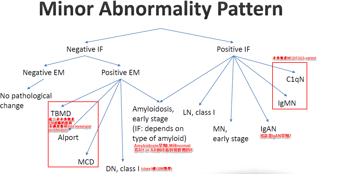
Minor Abnormality Pattern D/D:
                     Negative IF              Positive IF
                    ↙                        ↙              ↘
No pathological change   Amyloidosis,    C1qN         IgMN
                         LN class I,      ↘
                         early stage       DN, class I
Alport                                    early stage
MCD
5.Mesangioproliferative pattern#
- 定義
- Mesangial hypercellularity: ≥4 mesangial cells per mesangial area/glomerular tuft
- Oxford MEST-C 計分切點(切片需至少8顆腎絲球):M0(≤50% glomeruli)/M1(>50% glomeruli 具 mesangial hypercellularity >3 cells/area); E0(無)/E1(有 endocapillary hypercellularity); S0(無)/S1(有 segmental sclerosis); T0(≤25%)/T1(26-50%)/T2(>50% IFTA); C0(無)/C1(<25%)/C2(≥25% crescents)〔PMID 28341274〕
- 不過在hilar area有時候會≥4cells(所以hilar area(vascular pole)無法看cellularity)
- Increased mesangial matrix: width of mesangial matrix >2 mesangial cell nuclei in >25% of mesangial areas
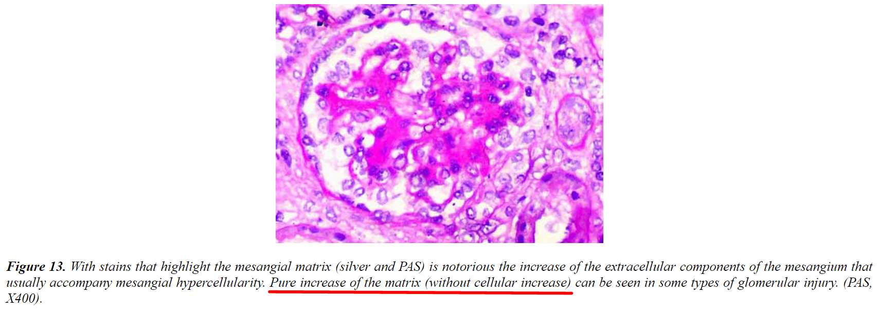
- Mesangioproliferative可以很輕微~很severe(甚至導致glomerular distorsion or marked lobulation of tuft(如此會有點像MPGN, 但大多不會這樣)); 大多mesangial matrix也增加(但也有isolated mesangial matrix增加=nodular pattern, 見下述)
- Mesangial hypercellularity: ≥4 mesangial cells per mesangial area/glomerular tuft
- D/D
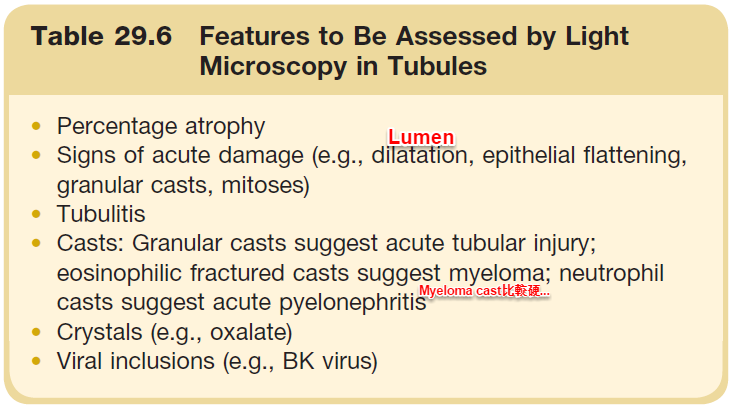
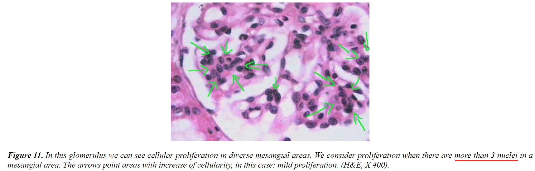
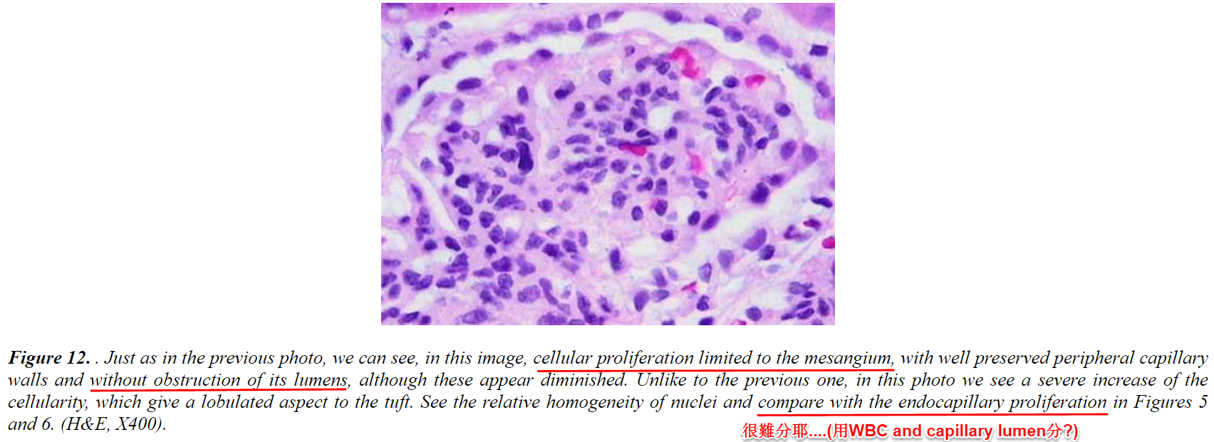
Figure 11. In this glomerulus we can see cellular proliferation in diverse mesangial areas. We consider proliferation when there are more than 3 nuclei in a mesangial area. The arrows point areas with increase of cellularity; in this case: mild proliferation. (H&E, X400).
Figure 12. Just as in the previous photo, we can see, in this image, cellular proliferation limited to the mesangium, with well preserved peripheral capillary walls and without obstruction of its lumens, although these appear diminished. Unlike to the previous one, in this photo we see a severe increase of the cellularity, which give a lobulated aspect to the tuft. See the relative homogeneity of nuclei and compare with the endocapillary proliferation in Figures 5 and 6. (H&E, X400).
Mesangioproliferative Pattern
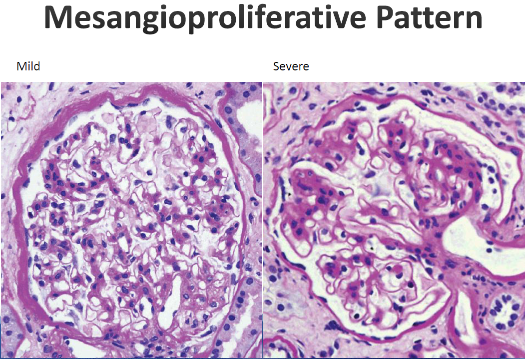
Mild | Severe
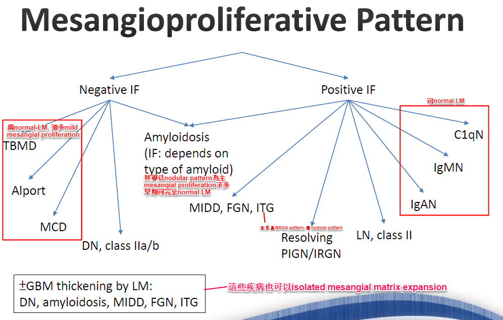
Mesangioproliferative Pattern - D/D#
                    Negative IF                        Positive IF
          ↙              ↘                   ↙                        ↘
Amyloidosis       (IF: depends on type   C1qN                         IgMN
(IF: depends        of amyloid)
on type of
amyloid)
                                          IgAN                    LN, class II
TBMD
Alport
MCD

DN, class IIa/b
↑GBM thickening by LM:               這些疾病有一些isolated mesangial matrix extension
DN, amyloidosis, MIDD, FGN, ITG
6.Nodular pattern#
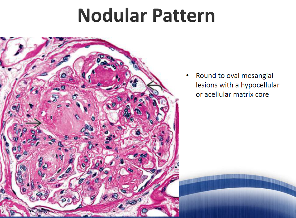
- 就是主要是mesangial matrix expansion(acellular or hypocellular)形成nodule, 不過可以多少有一些(hypocellular)mesangial proliferation
- 表示pathologic deposits of proteins or circulating substances(這些疾病也會堆積在GBM)
Nodular Pattern: Round to oval mesangial lesions with a hypocellular or acellular matrix core
- D/D
Organized glomerular electron-dense deposits algorithm:
                    Amyloid ←Congo red stain→ Nonamyloid
                   ↙                              ↘
Immunofluorescence for Ig LC & AA        Immunofluorescence for Ig
LC+              AA+                Non-Immunoglobulin-derived
   ↓               ↓                          ↓
AL amyloid    AA amyloid            Immunoglobulin-        Diabetic        Collagenofibrotic    Fibronectin
(8-15 nm)     (8-15 nm)              derived             sclerosis        glomerulopathy       glomerulopathy
 fibrils        fibrils            (5-20 nm fibrils)   (??)             (banded collagen     (10-15 nm fibrils)
                                        ↓                                    fibers)
                              Cryoglobulinemia    Systemic lupus    Immunotactoid      Fibrillary
                              (25-30 nm           erythematosus     glomerulopathy     glomerulonephritis
                               microtubules)      ("fingerprints")  (20-50 nm          (15-30 nm fibrils)
                               ⑤fingerprint        (fibrils)        microtubules)
Nodular Pattern D/D:
                Negative IF                          Positive IF
          ↙                ↘                    ↙              ↘
DN, class III            Amyloidosis          ITG           (?)
Idiopathic nodular       (IF depends on      ↘
glomerulosclerosis        type of amyloid)    MIDD          Fibrillary GN
Collagenofibrotic
glomerulopathy
- 各疾病特殊染色特徵見上方 Nodular Pattern 比較表
7.Membranous pattern#
- 僅MGN類(含LN, class V)
- Global thickening of GBM
Membranous Pattern: H&E: Global thickening of GBM
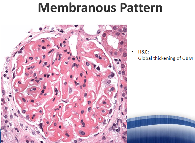
Figure 14. GBM reaction to the subepithelial deposits is seen as perpendicular projections to the GBM in its external part: "spikes", demonstrated here with silver stain (arrows). The immune deposits (no visible here) are between spikes. (Silver, X1000).
8.Necrotizing and crescentic pattern#
- Necrotizing指fibrinoid necrosis(會有GBM disruption, fibrin exudation and karyorrhexis(nuclei fragmentation))
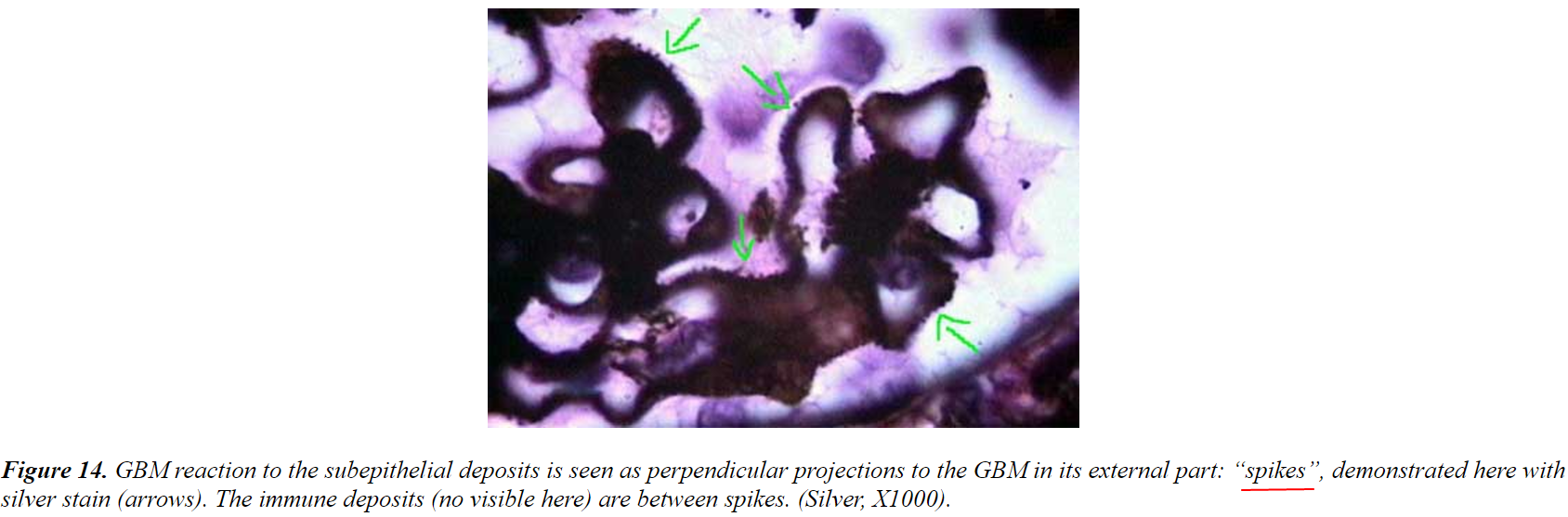
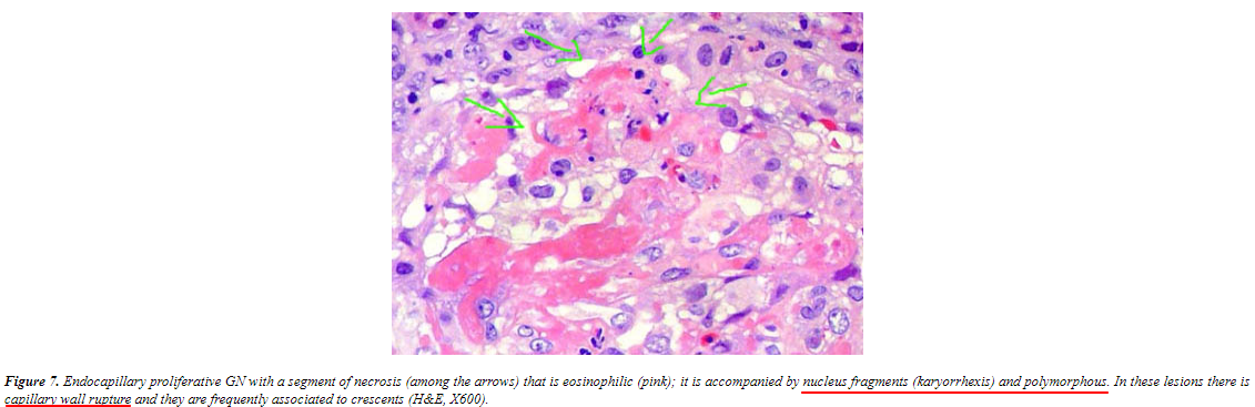
Figure 7. Endocapillary proliferative GN with a segment of necrosis (among the arrows) that is neutrophilic (pink); it is accompanied by nuclear fragments (karyorrhexis) and polymorphonuclear. In these lesions there is capillary wall rupture and they are frequently associated to a crescent. (H&E, X400).
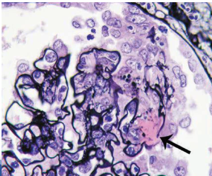
- Crescent指extracapillary proliferation
- Increased cellularity in the Bowman's space due to proliferation of the parietal epithelium or infiltration of inflammatory cells, fibroblasts(must be differentiated of podocyte proliferation(需要D/D collapsing FSGS, 因為collapsing FSGS的podocytes hyperplasia可以長得像crescent))
- 不過張富邦醫師的ppt說podocytes也可以包含在這些cells內, 所以和collapsing FSGS區分處在於crescent的細胞比較"雜"(因為很多種細胞嘛...)
- 定義:≧3 cell layers thick and cover ≥10% of the glomerular circumference(2018 ISN/RPS LN修訂對單一crescent的計數定義; 傳統教學常寫25%, AAV Berden的cellular/fibrocellular crescent亦無嚴格圈數門檻)
- 註:IgAN Oxford MEST-C 的 C 是以「有crescent的腎絲球比例」切分(C1<25%、C2≥25%), 與LN「單一crescent佔鮑氏囊≥10%」是不同軸的概念, 勿混用;Oxford 2016 更新:C 具預後預測力,建議加入 MEST 並以 MEST-C 報告〔PMID 28341274〕;LN 2018 修訂:active/chronic 改以修訂版 NIH activity index (0-24分,其中 fibrinoid necrosis 與 cellular/fibrocellular crescents 權重x2計分) 與 chronicity index (0-12分) 評估所有分型,並取消 IV-S/IV-G 次分型〔PMID 29459092〕
- 以前Crescentic GN定義為>50%的glomerulus都被involved, 但現在有crescent就算
- 原因是capillary wall的defect, 導致plasma products, fibrinogen進入Bowman's space, 進而導致fibrin formation(fibrinoid necrosis) and inflammatory cells influx(所以常伴隨endocapillary proliferation; 但若只有crescent 而沒有endocapillary proliferation:多為AAV or idiopathic crescent GN or anti-GBM disease), 進而導致cellular, fibrocellular and fibrous crescents(fibrous crescents多對immunosuppression沒有反應了)
- Crescent有cellular, fibrocellular, fibrous crescent
- 在AAV中fibrous crescent的定義是>90% of one crescent是fibrosis
- Increased cellularity in the Bowman's space due to proliferation of the parietal epithelium or infiltration of inflammatory cells, fibroblasts(must be differentiated of podocyte proliferation(需要D/D collapsing FSGS, 因為collapsing FSGS的podocytes hyperplasia可以長得像crescent))
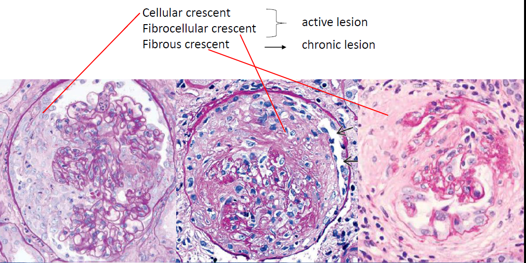
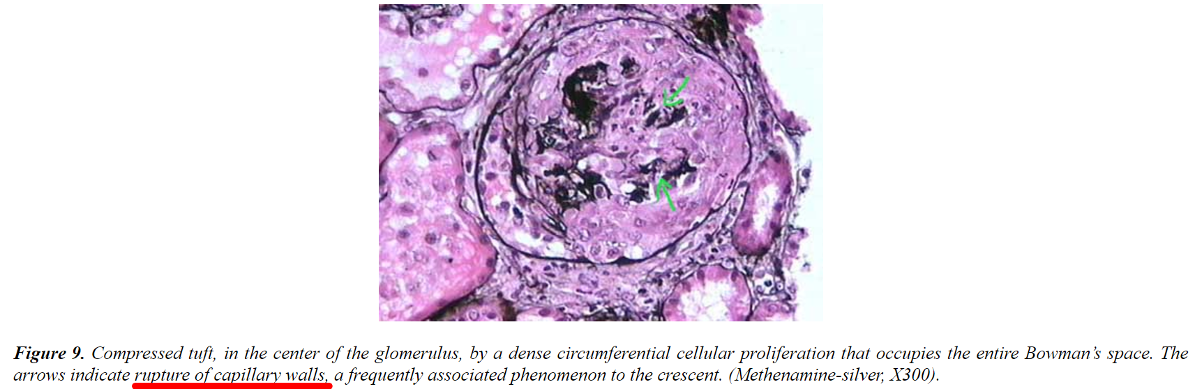
Cellular crescent → Fibrocellular crescent → active lesion → Fibrous crescent → chronic lesion
Figure 9. Compressed tuft in the center of the glomerulus, by a dense circumferential cellular proliferation that occupies the entire Bowman's space. The arrows indicate rupture of capillary walls, a frequently associated phenomenon to the crescent. (Methenamine-silver, X300).
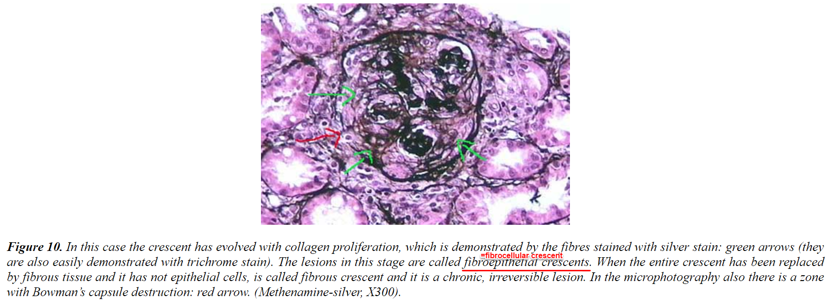
Figure 10. In this case the crescent has evolved with collagen proliferation, which is demonstrated by the fibres stained with silver stain: green arrows (they are also easily demonstrated with trichrome stain). The lesions in this stage are called fibrocellular crescents. When the entire crescent has been replaced by fibrous tissue and it has not epithelial cells, is called fibrous crescent and it is a chronic, irreversible lesion. In the microphotography also there is a zone with Bowman's capsule destruction: red arrow. (Methenamine-silver, X300).
- D/D主要是看immunofluorescence
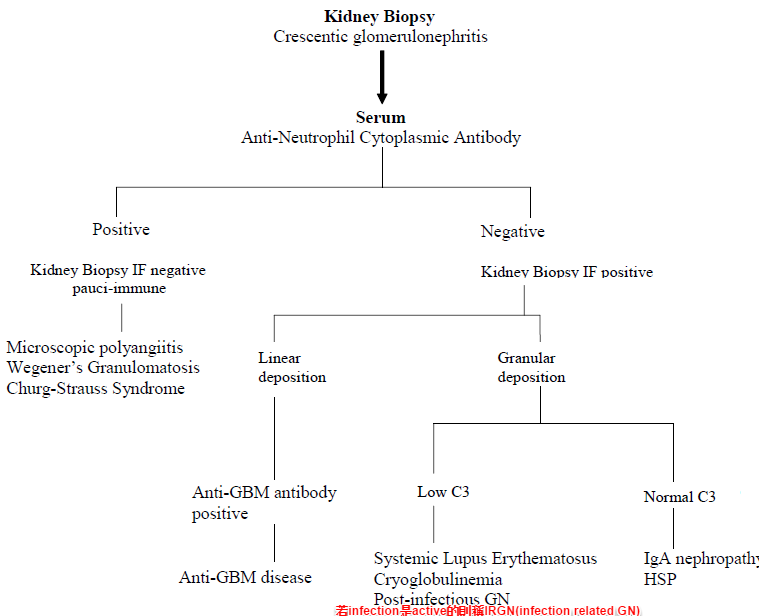
Kidney Biopsy - Crescentic glomerulonephritis Algorithm#
Kidney Biopsy
Crescentic glomerulonephritis
          ↓
       Serum
Anti-Neutrophil Cytoplasmic Antibody
    ↙                      ↘
Positive                  Negative
    ↓                          ↙        ↘
Kidney Biopsy IF negative   Kidney Biopsy IF positive
pauci immune                  ↙              ↘
                         Linear            Granular
                        deposition        deposition
Microscopic polyangiitis      ↙                  ↙          ↘
Wegener's Granulomatosis  Anti-GBM antibody  Low C3      Normal C3
Churg-Strauss Syndrome     positive              ↓             ↓
                          Anti-GBM disease   Systemic Lupus  IgA nephropathy;
                                             Erythematosus   HSP
                                             Cryoglobulinemia
                                             Post-infectious GN
                                             (Infection-related GN)
Necrotizing and Crescentic Pattern - IF Patterns#
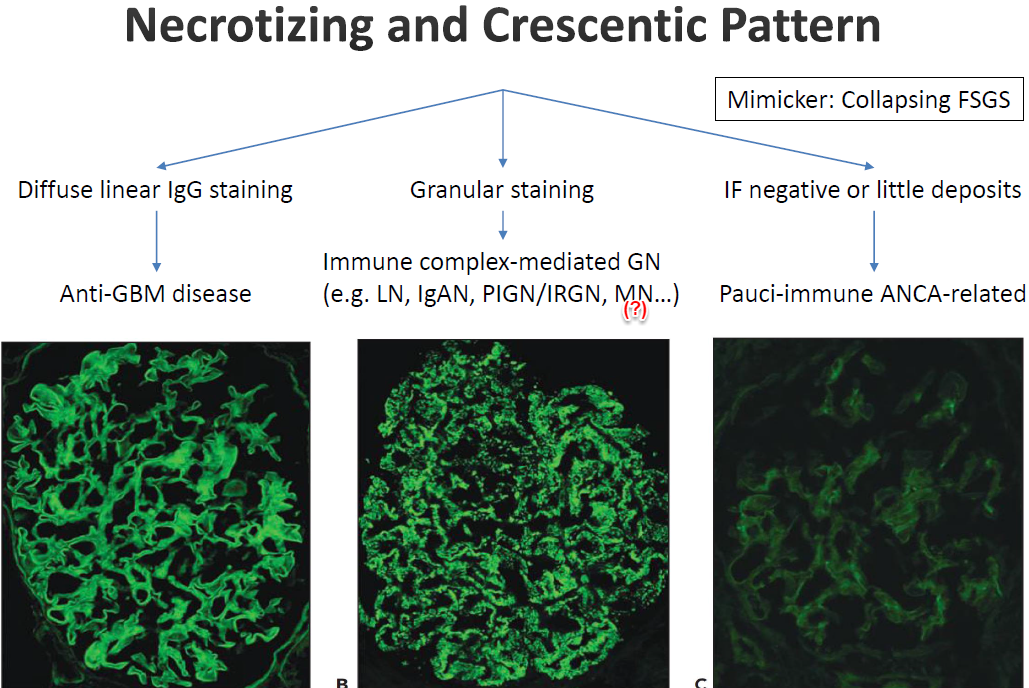
Diffuse linear IgG staining → Anti-GBM disease Granular staining → Immune complex-mediated GN (e.g. LN, IgAN, PIGN/IRGN, MN...) | Mimicker: Collapsing FSGS IF negative or little deposits → Pauci-immune ANCA-related
9.Endocapillary proliferative pattern#
- Endocapillary有兩個定義
- 有些僅定義intracapillary lumen, 有些定義為intracapillary lumen+mesangial area
- 一堆cells在capillary裡, usually accompanied with mesangial hypercellularity(所以一堆cells=endothelial, mesangial, WBCs增生(包含mononuclear cell, PMN...可染CLA(common leukocyte antigen), 不然LM比較好看的是PMN and eosinophils; lymphocytes不太好看...)
- Glomerulus變大(?)with變大的tufts, capillary lumens看不太到(壓扁 or 塞滿cells; 註:若單純只有mesangial cell proliferation and mesangial matrix expansion大多不會壓扁lumen)
- 導致glomerulus形成一個一個lobule(叫lobulation)
- Glomerulus變大(?)with變大的tufts, capillary lumens看不太到(壓扁 or 塞滿cells; 註:若單純只有mesangial cell proliferation and mesangial matrix expansion大多不會壓扁lumen)
- 這些疾病皆可MPGN pattern, 很嚴重吃破capillary lumen的話會fibrinoid necrosis, crescent GN
- No duplication of GBM(未必吧...)
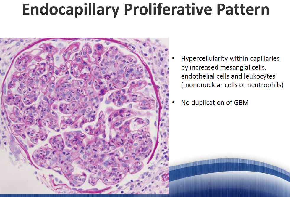
Endocapillary Proliferative Pattern: Hypercellularity within capillaries by increased mesangial cells, endothelial cells and leukocytes (mononuclear cells or neutrophils). No duplication of GBM.
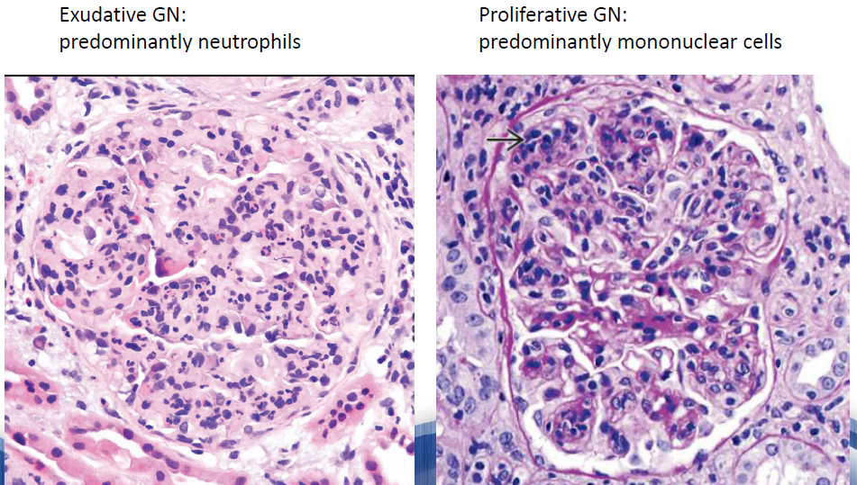
Exudative GN: predominantly neutrophils | Proliferative GN: predominantly mononuclear cells
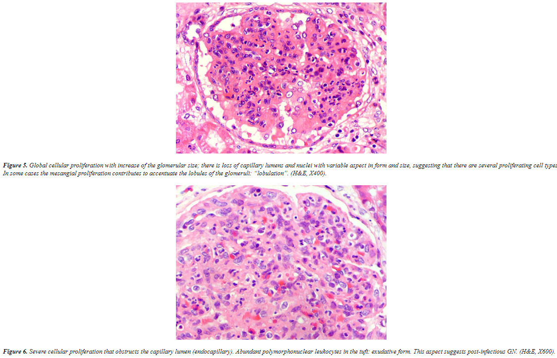
Figure 5. Global cellular proliferation with increase of the glomerular size: there is loss of capillary lumens and nuclei with variable aspects in form and size, suggesting that there are several proliferating cell types. In some cases the mesangial proliferation contributes to accentuate the lobules of the glomerulus "lobulation". (H&E, X40).
Figure 6. Severe cellular proliferation that obstructs the capillary lumen (endocapillary). Abundant polymorphonuclear leukocytes in the tuft: exudative form. This aspect suggests post-infectious GN. (H&E, X800).
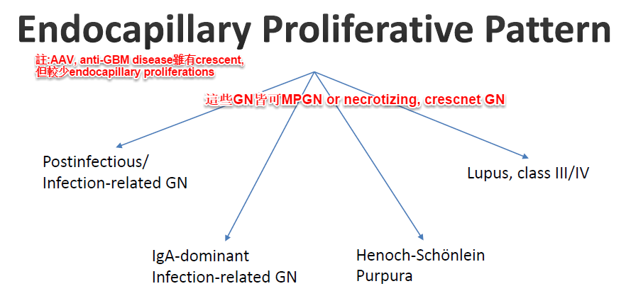
- D/D
Endocapillary Proliferative Pattern - D/D#
   (↑)AAV, anti-GBM disease的 crescent;
   其他endocapillary proliferations
              ↙(+)如↑MPGN or necrotizing, crescent GN
         ↙
Postinfectious/               Lupus, class III/IV
Infection-related GN
         ↘
     IgA-dominant
     Infection-related GN
                   Henoch-Schönlein Purpura
10.Membranoproliferative pattern(又叫Mesangiocapillary pattern)#
- 會mesangial hypercellularity+mesangial matrix expansion(前2者導致lobulation appearance)+endocapillary proliferation+double contour due to mesangial interposition(tram track)(尤其silver stain可以明顯看出)
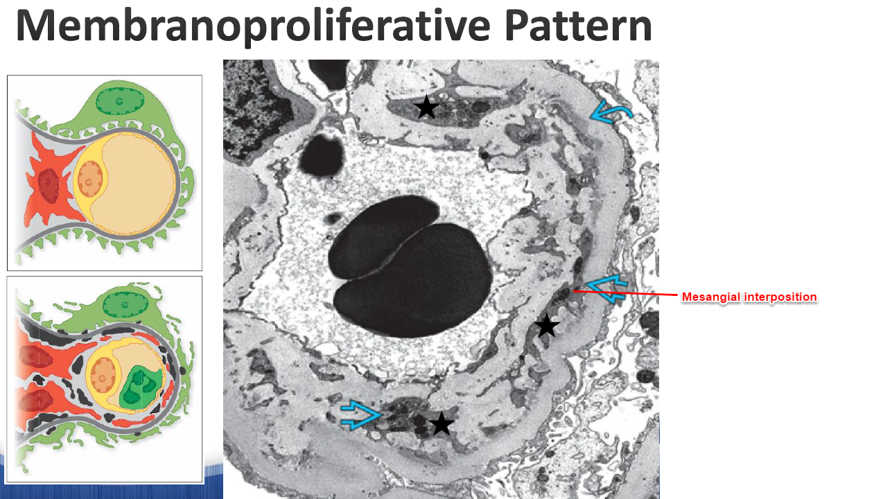
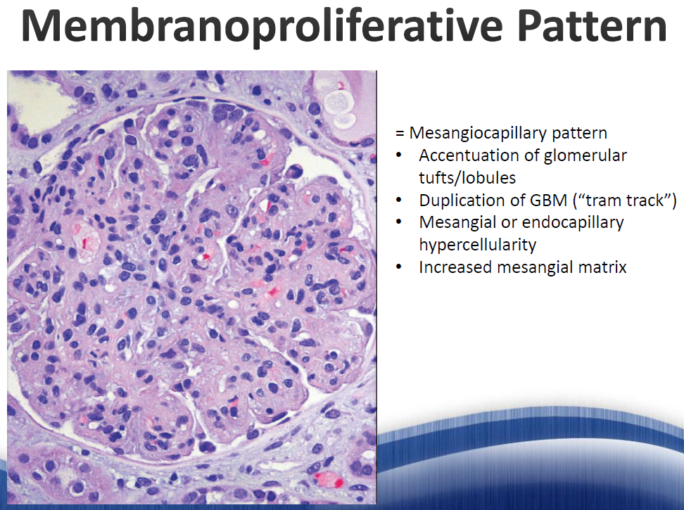
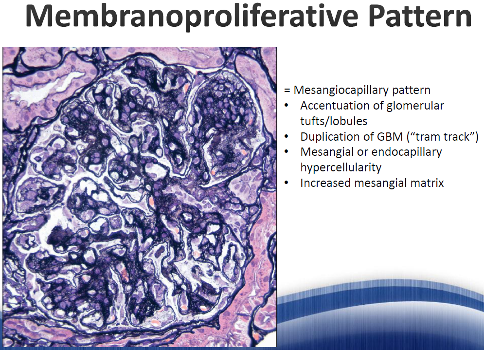
Membranoproliferative Pattern = Mesangiocapillary pattern
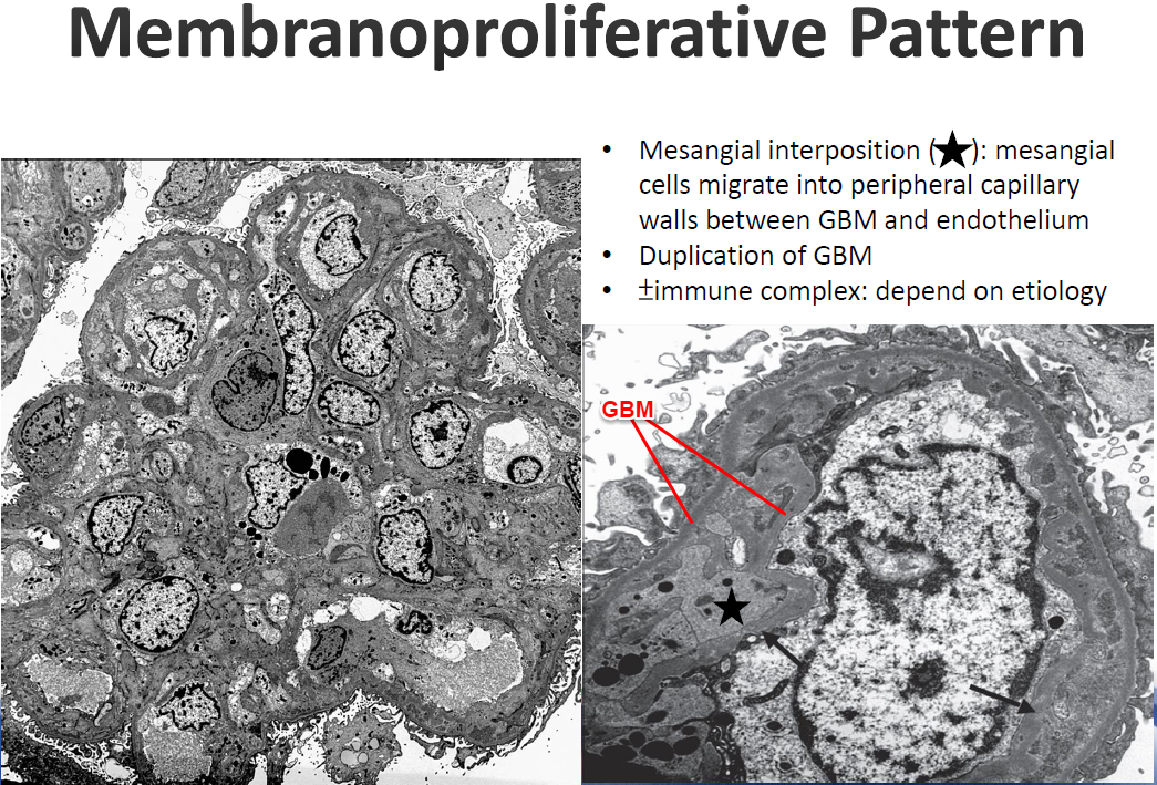
- Accentuation of glomerular tufts/lobules
- Duplication of GBM ("tram track")
- Mesangial interposition (★): mesangial cells migrate into peripheral capillary walls between GBM and endothelium (±immune complex: depend on etiology)
- Mesangial or endocapillary hypercellularity
- Increased mesangial matrix
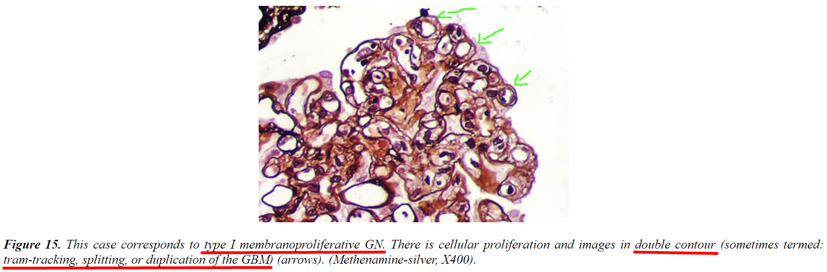
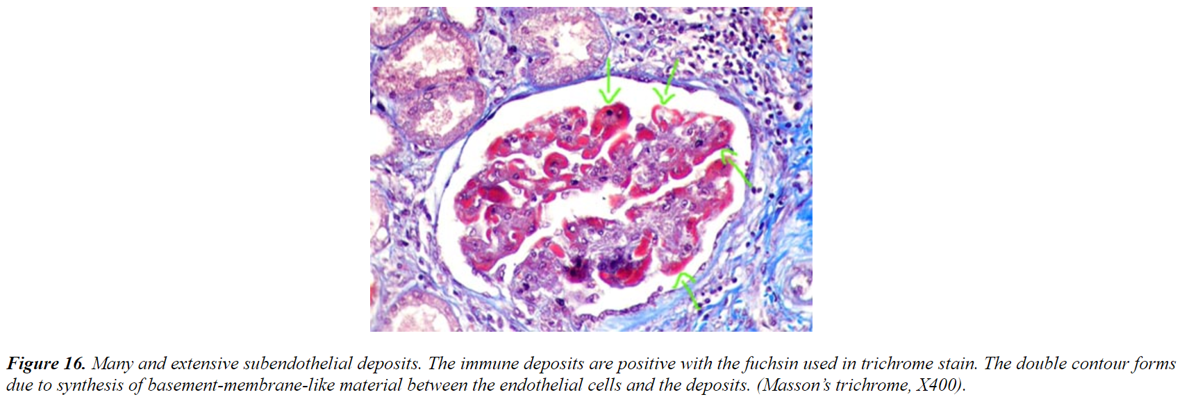
Figure 15. This case corresponds to type I membranoproliferative GN. There is cellular proliferation and images in double contour (sometimes termed: tram-tracking, splitting, or duplication of the GBM) (arrows). (Methenamine-silver, X400).
Figure 16. Many and extensive subendothelial deposits. The immune deposits are positive with the fuchsin used in trichrome stain. The double contour forms due to synthesis of basement-membrane-like material between the endothelial cells and the deposits. (Masson's trichrome, X400).
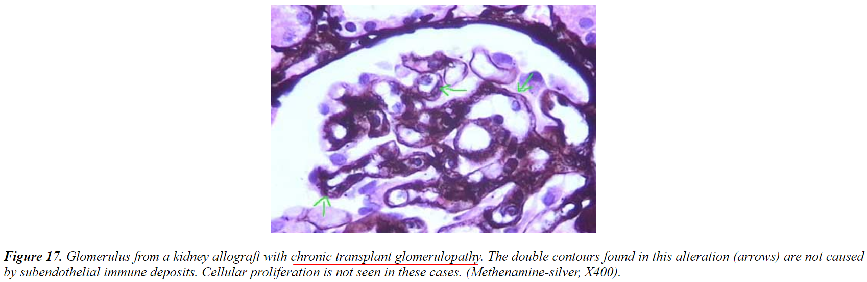
Figure 17. Glomerulus from a kidney allograft with chronic transplant glomerulopathy. The double contours found in this alteration (arrows) are not caused by subendothelial immune deposits. Cellular proliferation is not seen in these cases. (Methenamine-silver, X400).
-
機轉與病理特徵見上方條列
-
D/D
Membranoproliferative Pattern - D/D#
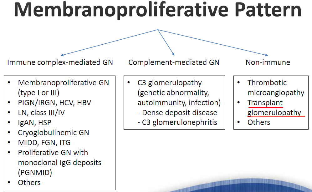
Immune complex-mediated GN        Complement-mediated GN       Non-immune
- Membranoproliferative GN        - C3 glomerulopathy           - Thrombotic
  (type I or III)                   (genetic abnormality,          microangiopathy
- PIGN/IRGN, HCV, HBV              autoimmunity, infection)     - Transplant
- LN, class III/IV                  - Dense deposit disease        glomerulopathy
- IgAN, HSP                         - C3 glomerulonephritis      - Others
- Cryoglobulinemic GN
- MIDD, FGN, ITG
- Proliferative GN with
  monoclonal IgG deposits
  (PGNMID)
- Others
11.Focal segmental sclerosing pattern(就是FSGS...)(should exclude other GN first)#
- Focal (only in some glomeruli) or diffuse (in all or almost all the glomeruli)
- Segmental (only a part of the glomerulus) or global (entire glomerulus)
- Sclerosis的定義是distorted capillary tufts with obliteration of lumen by increased matrix, glassy hyaline material or foam cells; +/- adhesions to Bowman's capsule
- 註:Global sclerosis(FGGS):may be ischemic change; could be aging change; 也可以是rests of a crescent or remaining fragments of tuft fibrinoid necrosis or immune deposits in not completely sclerosed tufts(=過去傷害的殘骸)
- A characteristic pattern of retraction and collapse of capillary tuft (PAS(+)) surrounded by fibrotic tissue (PAS(-))(as below)
- 註:Global sclerosis(FGGS):may be ischemic change; could be aging change; 也可以是rests of a crescent or remaining fragments of tuft fibrinoid necrosis or immune deposits in not completely sclerosed tufts(=過去傷害的殘骸)
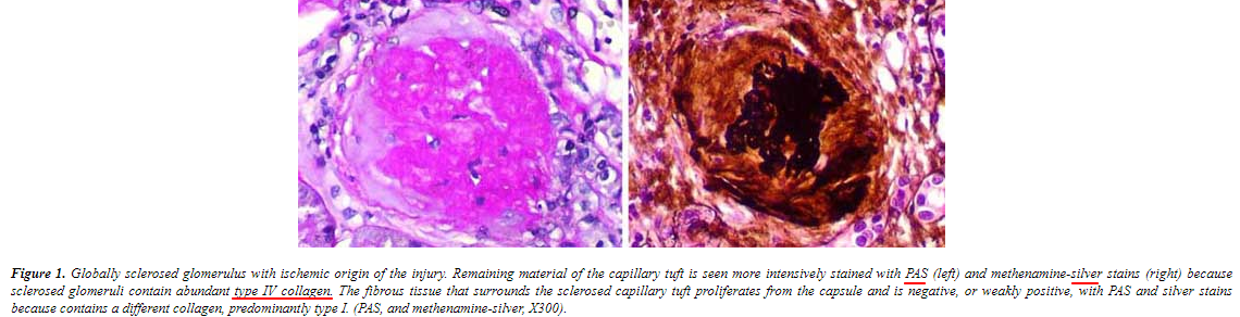
Figure 3. Globally sclerosed glomerulus with ischemic origin of the lesion. Remaining material of the capillary tuft is seen more compacted (stained with H&E left) and methenamine-silver stain right. Tightly because the fibrous tissue that surrounds the sclerosed capillary tuft proliferates from the capsule and a negative, or weakly positive, H&E and silver stain X40). In other cases fibrous tissue is a stiff collagen, predominantly type I (V), and methenamine-silver stain X40).
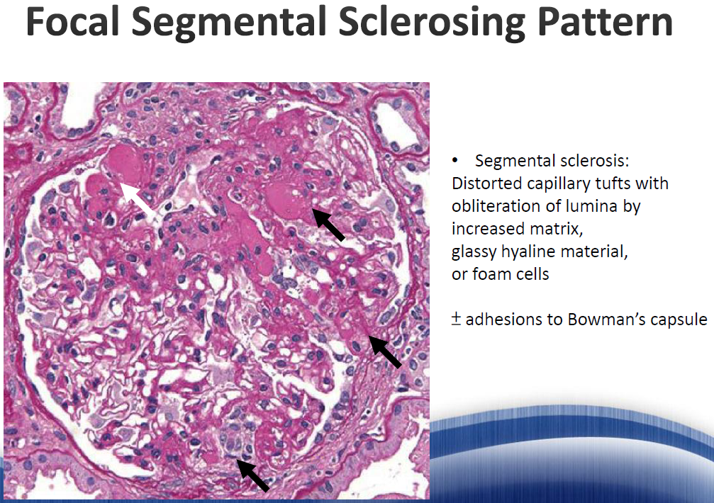
Focal Segmental Sclerosing Pattern
- Segmental sclerosis: Distorted capillary tufts with obliteration of lumina by increased matrix, glassy hyaline material, or foam cells
- ± adhesions to Bowman's capsule
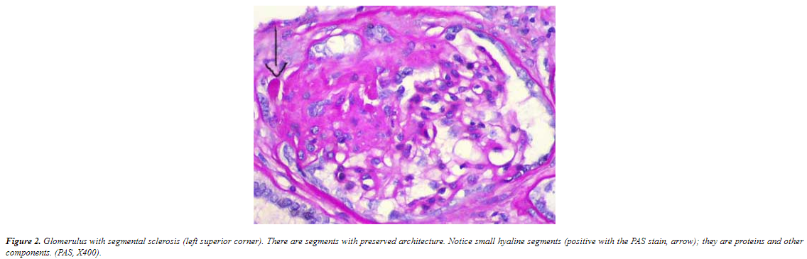
Figure 2. Glomerulus with segmental sclerosis (left superior corner). There are segments with preserved architecture. Notice small hyaline segments (positive with the PAS stain: arrow); they are proteins and other components. (PAS, X400).
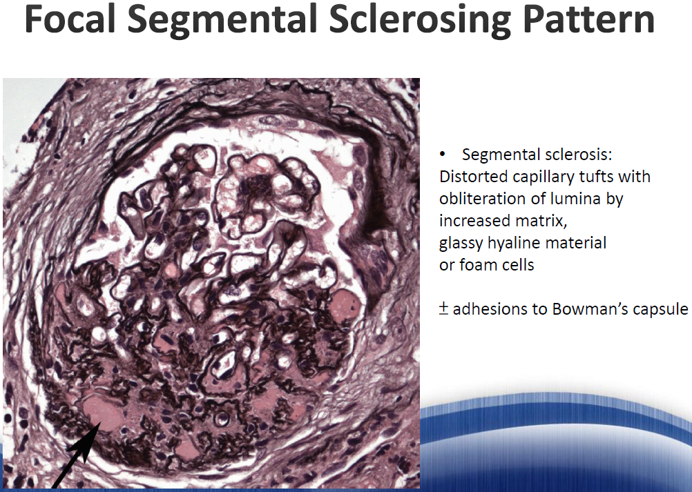
Focal Segmental Sclerosing Pattern (silver stain → 見上方定義)
- Classification
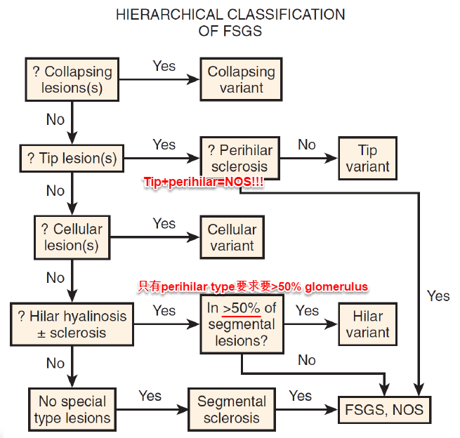
Hierarchical Classification of FSGS (Columbia 五型:collapsing, tip, cellular, perihilar, NOS;Tip 緩解率最高59.5%、預後最佳,Collapsing 緩解率最低24.5%、43.4%腎衰竭、預後最差)〔PMID 38596270〕#
? Collapsing lesion(s) → Yes → Collapsing variant
                ↓ No
? Tip lesion(s) → Yes → ? Perihilar sclerosis → No → Tip variant
                ↓ No       Tip+perihilar:NOS(非特異型)!
? Cellular lesion(s) → Yes → Cellular variant
                ↓ No   且:perihilar type要≥50% glomerulus
? Hilar hyalinosis ± sclerosis → Yes → In >50% of segmental lesions? → Yes → Hilar variant
                ↓ No                          ↓ No
No special type lesions → Yes → Segmental sclerosis → Yes → FSGS, NOS
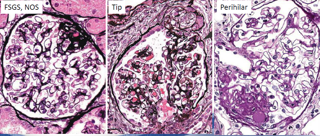
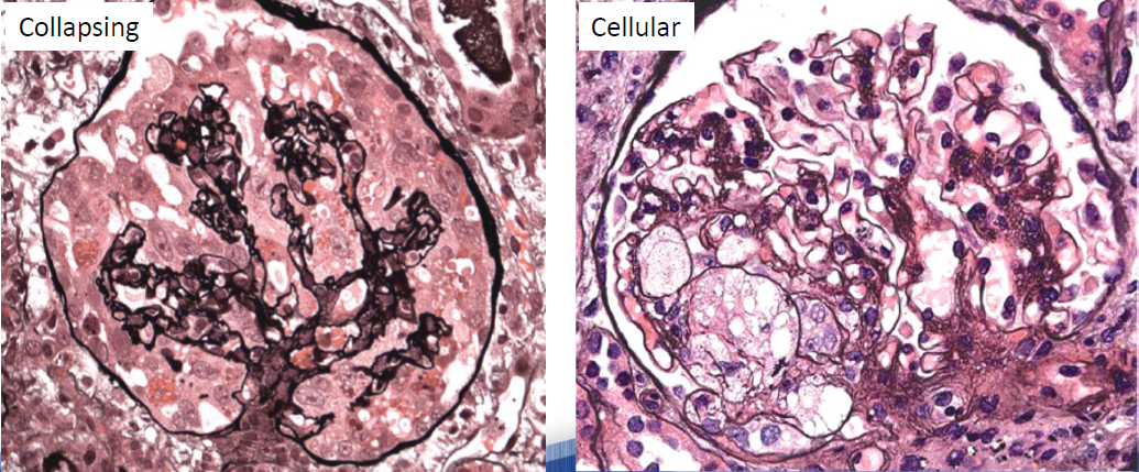
FSGS, NOS | Tip | Perihilar | Collapsing | Cellular
- Others
- Fabry's disease
Others - Fabry Nephropathy#
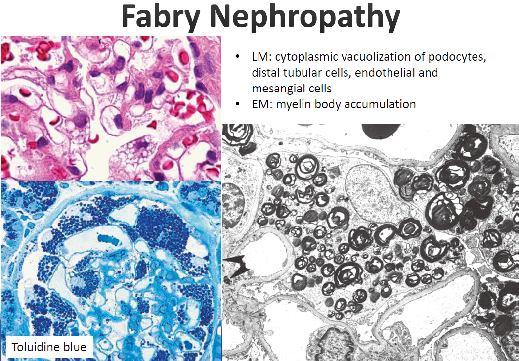
Fabry Nephropathy
-
LM: cytoplasmic vacuolization of podocytes, distal tubular cells, endothelial and mesangial cells
-
EM: myelin body accumulation
-
Toluidine blue stain
-
HTN nephropathy/HTN effects/malignant HTN
HTN - Hypertensive Nephropathy#
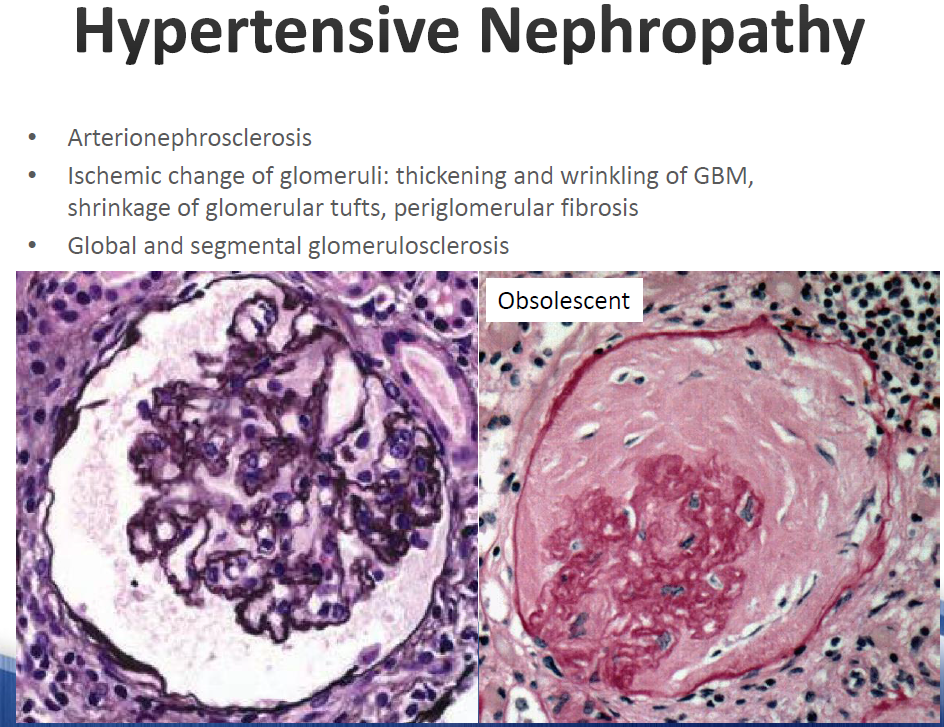
Hypertensive Nephropathy
- Arterionephrosclerosis
- Ischemic change of glomeruli: thickening and wrinkling of GBM, shrinkage of glomerular tufts, periglomerular fibrosis
- Global and segmental glomerulosclerosis
- Obsolescent glomerulus (有時候可見Bowman's capsule附近有fibrosis)
Essential Hypertension#
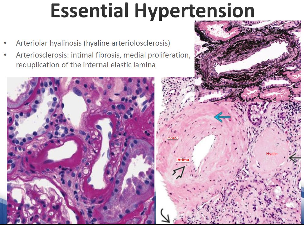
- 可以是periglomerular fibrosis(interstitial nephritis久了) or (subcapsular fibrosis due to HTN)
- 可用Silver stain區分上述二者, Bowman's capsule會比fibrosis厚
- Essential Hypertension
- Arteriolar hyalinosis (hyaline arteriolosclerosis)
- Arteriosclerosis: intimal fibrosis, medial proliferation, reduplication of the internal elastic lamina
Malignant or Accelerated Hypertension#
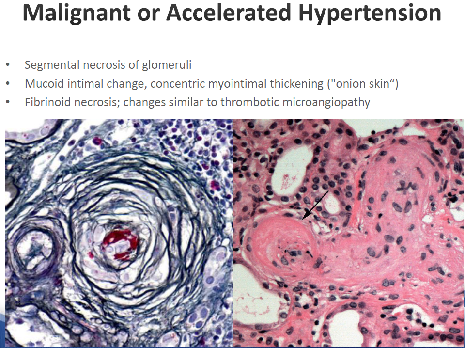
Malignant or Accelerated Hypertension
-
Segmental necrosis of glomeruli
-
Mucoid intimal change, concentric myointimal thickening ("onion skin")
-
Fibrinoid necrosis; changes similar to thrombotic microangiopathy
-
Cast nephropathy
Cast Nephropathy - Light Chain Cast Nephropathy#
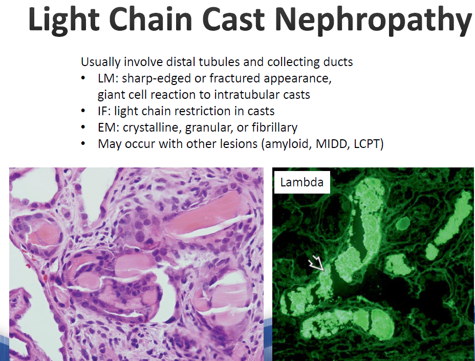
Light Chain Cast Nephropathy
-
Usually involve distal tubules and collecting ducts
-
LM: sharp-edged or fractured appearance, giant cell reaction to intratubular casts
-
IF: light chain restriction in casts
-
EM: crystalline, granular, or fibrillary
-
May occur with other lesions (amyloid, MIDD, LCPT)
-
Lambda stain (IF)
-
Light chain proximal tubulopathy (Fanconi syndrome)
Light Chain Proximal Tubulopathy#
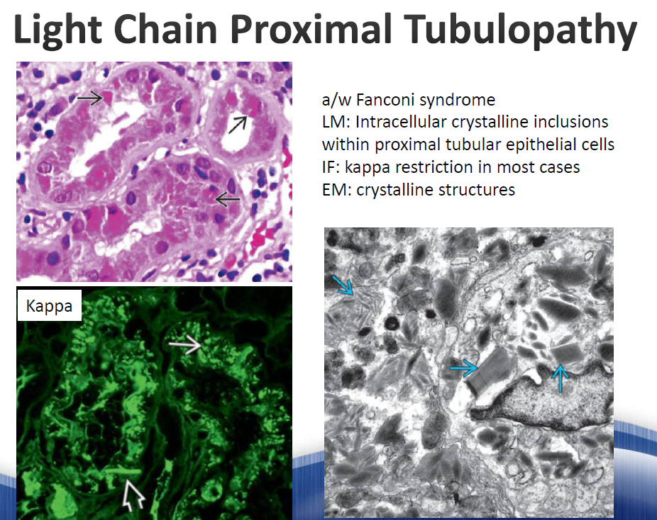
Light Chain Proximal Tubulopathy
- a/w Fanconi syndrome
- LM: Intracellular crystalline inclusions within proximal tubular epithelial cells
- IF: kappa restriction in most cases
- EM: crystalline structures
12.Tubules/Interstitial#
- 可看Masson trichrome stain, 正常proximal tubules是一個和一個貼在一起, aging/fibrosis的話tubules才會分比較開
- Proximal tubules比distal tubules在HE stain下較tall, eosinophilic, 且有brush border(表面的microvilli)
- Proteinuria/nephrotic syndrome久了因為proximal tubules會吸收很多proteins, 所以tubular cells會變得有點foamy(vacuolization; D/D:CNI use, mannitol, IVIG, dextran, contrast)
- Tubular atrophy, tubules會扁掉, lumen變小, basement membrane變厚
- Medulla本來interstitial就比較多所以難說interstitial fibrosis, 除非>30-40%; cortex則應<10%
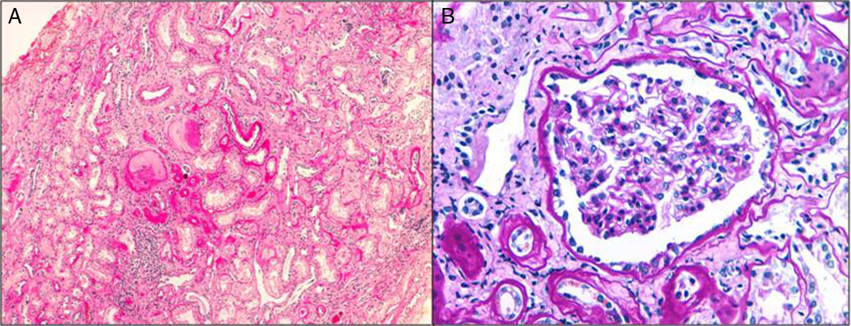
(A) Interstitial fibrosis, focal tubular atrophy, glomerular sclerosis and mild chronic inflammatory infiltrate (H–E stain, 4×) (B) Glomerulus without optical morphological alterations (PAS staining, 20×)
- 註:PAS stain蠻好看Bowman's capsule, mesangial matrix, basement membrane(BM)
- Chronic tubular atrophy可以thyroidization(dilated tubular segments with eosinophilic proteinaceous casts and thin epithelial lining)(尤其chronic PN, 不過not specific)
Thyroidization of Tubules#
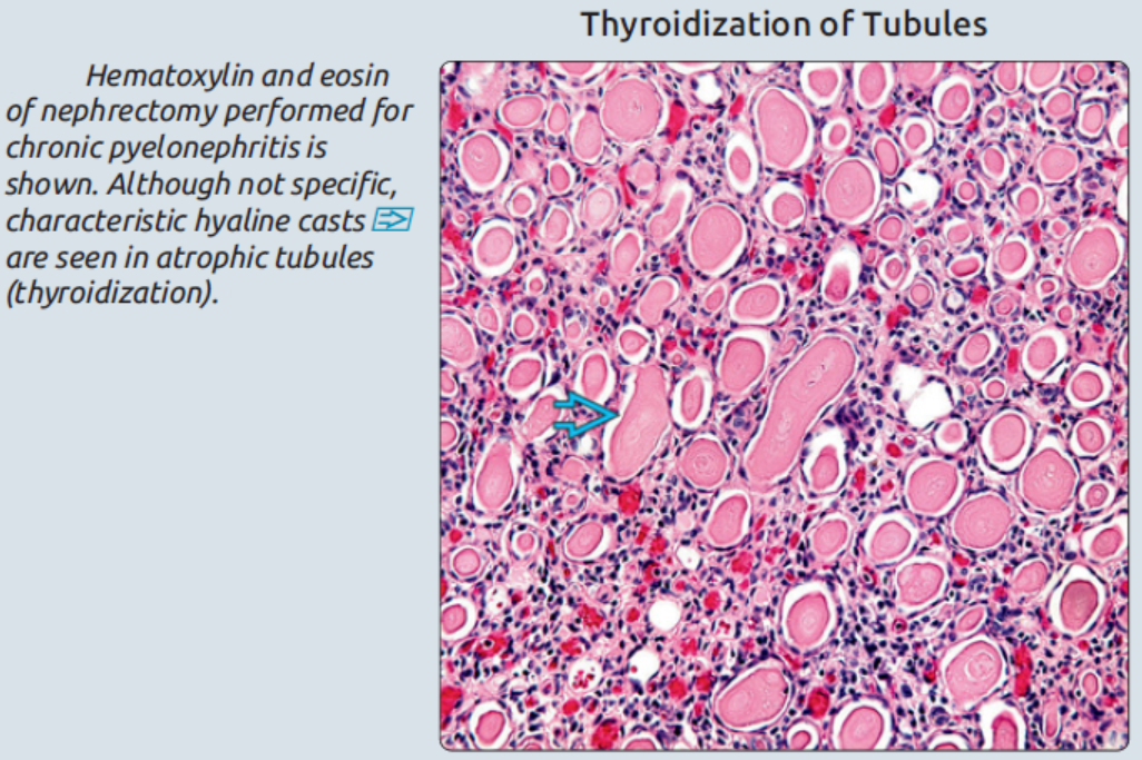
Hematoxylin and eosin of nephrectomy performed for chronic pyelonephritis is shown. Although not specific, characteristic hyaline casts are seen in atrophic tubules (thyroidization).
- AIN
- Glomerulus, BV
- 不太有變化
- Interstitium
- 會edema, WBCs infiltration(eosinophils大多在早期出現, 接下來以lymphocytes為主(主要是CD4(+) T cells, 不過 CD8(+)T cells, B lymphocytes, plasma cells, NK cells, macrophages也有); 可能會有granuloma; 第二週(含)以後fibrosis就開始出現了!
- 註:但NSAID的AIN以CD8(+) T cells為主且少eosinophils
- 似乎involve cortex more than medulla(同Chinese herb nephropathy)
- 會edema, WBCs infiltration(eosinophils大多在早期出現, 接下來以lymphocytes為主(主要是CD4(+) T cells, 不過 CD8(+)T cells, B lymphocytes, plasma cells, NK cells, macrophages也有); 可能會有granuloma; 第二週(含)以後fibrosis就開始出現了!
- Tubules
- 比較嚴重的會有tubulitis
- Immunofluorescence(IF)有時候可見immunocomplex and complements沉積在TBM, 但IF(-)比較常見
- Glomerulus, BV
- CIN/cTIN
- Glomerulus
- 原則上不太有變化, 但有periglomerular fibrosis(D/D HTN的subcapsular fibrosis)
- CTIN久了會glomerular sclerosis
- BV
- Variable fibrointimal thickening
- 以IFTA為主, cellular infiltrations比AIN少, tubular BM會變厚, 可以有anti-TBM的IgG, C3(IF)
- Glomerulus
- ATN
- Glomerulus
- 不太有變化, 不過最近發現似乎有一些podocytes change(EM?)
- PCT
- Loss of apical brush border
- Cell detachment-->到DCT變成epithelial casts
- The sloughed tubular cells, brush border vesicle remnants, and cellular debris in combination with T-H protein form the classical "muddy-brown granular" casts
- Lumen dilation
- Glomerulus
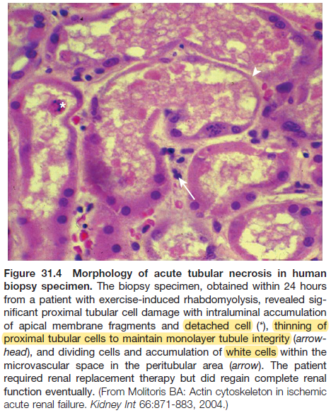
Figure 31.4 Morphology of acute tubular necrosis in human biopsy specimen. The biopsy specimen, obtained within 24 hours from a patient with exercise-induced rhabdomyolysis, revealed significant proximal tubular cell damage with intraluminal accumulation of apical membrane fragments and detached cell (), thinning of proximal tubular cells to maintain monolayer tubule integrity (arrowhead), and dividing cells and accumulation of white cells within the microvascular space in the peritubular area (arrow). The patient required renal replacement therapy but did regain complete renal function eventually. (From Molitoris BA: Actin cytoskeleton in ischemic acute renal failure. Kidney Int 66:871-883, 2004.)*
- For aristolochic acid nephropathy(Chinese herb nephropathy)
- Early lesions have ATN with interstitial myxoid change and scattered mononuclear cell infiltrates(仍然比一般的IN少inflammation)
- Late interstitial fibrosis tends to be paucicellular and most pronounced in outer cortex
- 少有glomerulus change, 除非晚期
13.Vessels: vasculitis and arteriosclerosis#
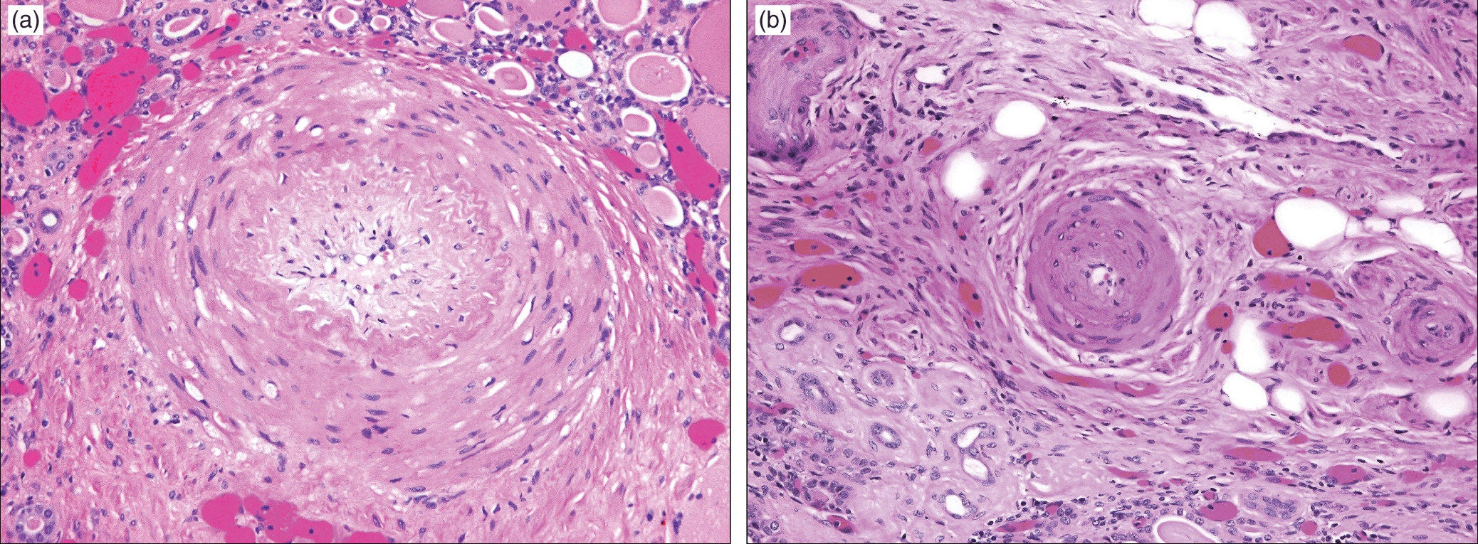
(A) Typical pattern of arteriopathy in ESRD with marked luminal narrowing, intimal fibrosis, and medial smooth muscle hyperplasia (B) A small muscular artery with typical arteriopathy of ESRD with marked luminal narrowing, medial smooth muscle hyperplasia, and periarterial fibrosis
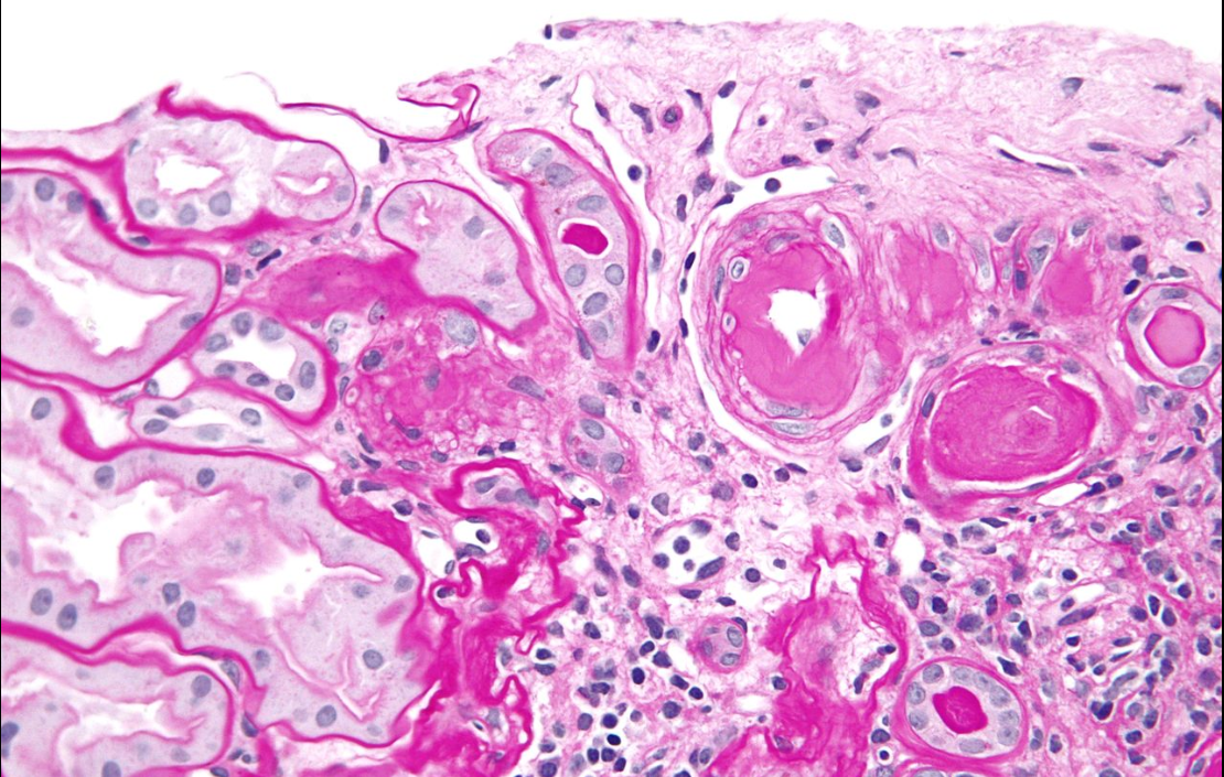
- PAS: hyaline arteriosclerosis